|
Roll Overs:
#1
#2
#3
#4
#5
#6
|
|
|
Copyright (c) Steve Brittenham.
|
What is a thin section? What do all the colors mean?
|
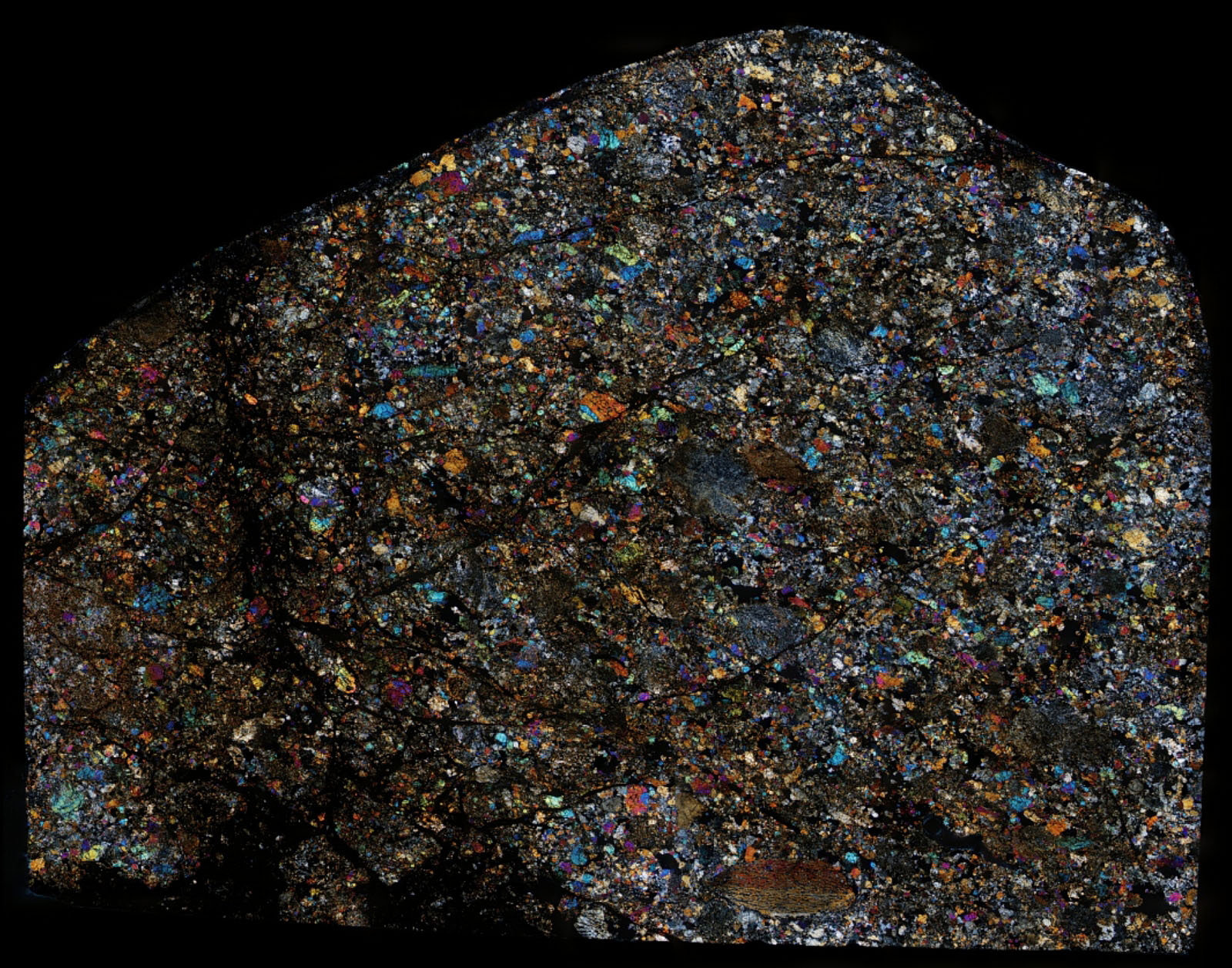
Thin section. L6
TKW 37 kg. Observed fall 26 April 1803, Normandy, north-west of Paris, France.
    
Steve writes:
Since there are already nice examples of fusion crusted L�Aigle pieces on MPOD, I thought I�d offer instead a high resolution thin section scan to show what this historic meteorite looks like under a microscope.
Photo 1 is a low resolution view of a 2.5 gigapixel (56835 by 44510 pixel) mosaic built from 208 individual 18 Mpixel captures. You can pan around and zoom in and out of the entire full-res image here
(If you haven�t used GigaPan before, go full screen by clicking on the diagonal arrows at the right of the image, then pan around with your mouse and zoom in and out with its scroll wheel.)
The thin section�s fusion crust and shock veins are more obvious in Photo 2�s low resolution white light image, Photo 3�s low resolution animated overlay, and in the full resolution image here
Photo 4�s xpol and brightfield comparison shows one of the few well-defined chondrules in this thin section. But there are many other interesting areas to view (Photo 5 is an example with a mix of features, some of which are bisected by a shock vein).
For those interested in how these images were created, I got into full slide thin section imaging when I was asked to do one for an unusual rock. After almost three hours of manually positioning the stage, I had both the 324 pictures necessary for a final stitch, and a newfound commitment never to do that again!
So I cobbled together about $85 worth of electronics to control my microscope stage and camera, then spent the next several weeks programming, debugging, optimizing, and calibrating everything. Photo 6 shows the current setup (yup, that�s dishwasher drain hose connecting the stepper motors to the microscope stage!).
The joystick is used to specify two opposite corners of an area to be imaged, then the controller automatically positions and triggers successive photo captures with appropriate horizontal and vertical overlap. Even large thin sections can be imaged in under 15 minutes, with another 15 for the mosaicking software to process them. (But because of the large files sizes, any sharpening or HDR pre-processing of the image set adds another 90 minutes each!)
Admittedly, I probably could have manually imaged all of my thin sections in the time it took to develop the system; but between my attention deficit and a lack of any real patience, this was the better solution! |
Click to view larger photos
#1
#2
#3
#4
#5
#6
|
Found at the arrow (green or red) on the map below
|
|
| |
John Lutzon
8/18/2020 7:23:33 PM |
Maybe not You, but I wish I was your next door neighbor.Beautiful work/results, great resourcefulness. |
Benjamin P. Sun
8/18/2020 6:04:08 AM |
Nice setup! Great pics |
Bernd Pauli
8/18/2020 3:57:12 AM |
As for photo #4, I've seldom seen a BO (= barred olivine) chondrule that is so severely elongated or compressed. Thanks for sharing these thin section impressions! |
| |
|